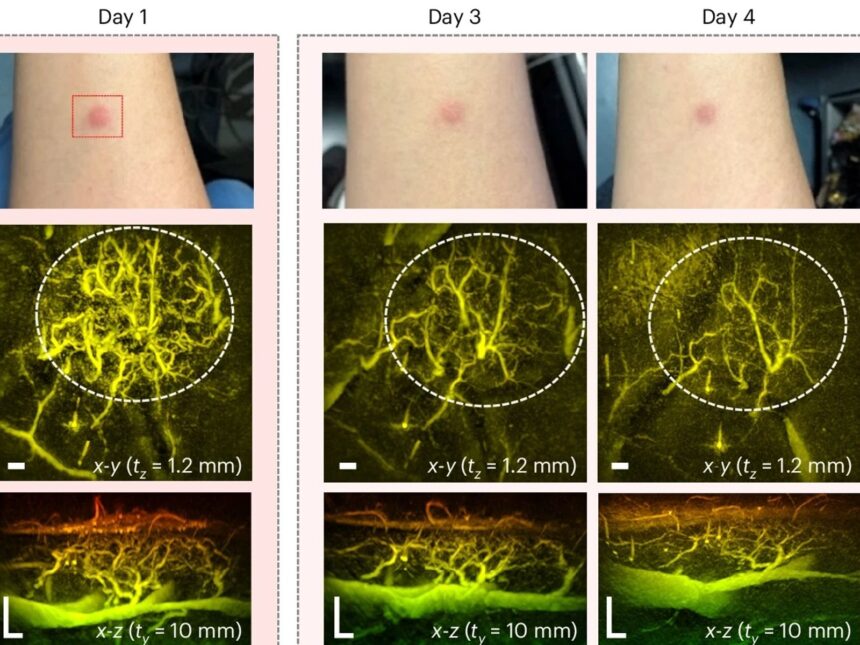
The technology, created by researchers at University College London (UCL), is a revolutionary new tool for the diagnosis and treatment of conditions like diabetes, arthritis, and some types of cancer since it is especially good at visualizing blood arteries.
It employs a method known as Photoaccoustic Tomography (PAT), which combines laser light with the ultrasonic waves it produces in specific tissues to create a real-time, three-dimensional picture of human biology.
Although the method was developed over 20 years ago, earlier iterations took many seconds or even minutes to capture an image.
The researchers at UCL have lowered that time to a second or less.
With their discovery, they intend to develop a handheld scanner that may be used in clinics on a regular basis to replace expensive imaging machines like MRIs and dangerous X-rays.
Professor Paul Beard, a medical physicist at UCL who participated in the study, said, “These technological advancements make the system suitable for clinical use for the first time, allowing us to look at aspects of human biology and disease that we haven’t been able to look at before.”


